 مد بوک | جامع ترین آرشیو کتاب های پزشکی
Renowned for its superb illustrations and highly practical information, the third volume of this classic reference reflects the very latest in state-of-the-art imaging technology. Together with Volumes 1 and 2, this compact and portable book provides a highly specialized navigational tool for clinicians seeking to master the ability to recognize anatomical structures and accurately interpret CT and MR images.
Highlights of Volume 3:
New CT and MR images of the highest quality
Didactic organization using two-page units, with radiographs on one page and full-color illustrations on the next
Concise, easy-to-read labeling on all figures
Color-coded, schematic diagrams that indicate the level of each section
Sectional enlargements for detailed classification of the anatomical structure
Comprehensive, compact, and portable, this popular book is ideal for use in both the classroom and clinical setting.
Contents
Section 1: Upper Extremity
Section 2: Lower Extremity
Section 3: Spinal Column
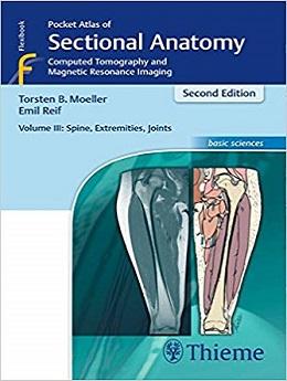
دانلود کتاب اطلس آناتومی مقطعی، جلد ۳: ستون فقرات، پایانگاه، مفاصل: توموگرافی کامپیوتری و تصویربرداری رزونانس مغناطیسی Pocket Atlas of Sectional Anatomy, Volume III: Spine, Extremities, Joints: Computed Tomography and Magnetic Resonance Imaging, 2ed
| نویسنده | orsten Bert Möller , Emil Reif |
| ناشر | TPS; 2nd edition |
| سال انتشار | 2017 |
| زبان کتاب | انگلیسی |
| نوع فایل | |
| تعداد صفحات | 482 |
| (ISBN) شابک | 3131431725 |
| حجم فایل | 43 MB |
168,000 تومان


محصولات مرتبط









نقد و بررسی وجود ندارد.